Couse

放射線医学

Course introduction

講座紹介

器官・形態領域
放射線医学(旧名称:放射線医学講座)

人体の様々な臓器における新たな技術及び撮像装置を用いた画像診断に関する研究や、新たな画像診断の開発を目指した研究を行っている。

【講座問い合わせ先】
器官・形態領域 放射線医学 〒791-0295愛媛県東温市志津川454
TEL:089-960-5371 / FAX:089-960-5375
公式HP https://www.m.ehime-u.ac.jp/school/radiology/

Course introduction

研究内容

  • 頭部放射線診断

機械学習を用いたMRI画像解析に関する研究、MRI画像による脳腫瘍の経過や予後予測に関する研究

  • 胸部放射線診断

・人工知能による診断や画像再構成に関する研究

・MRIによる画像情報の定量化、撮影の高速化に関する研究

  • 乳腺放射線診断

乳腺病変の診断精度向上を目的とする研究、画像による乳癌の免疫組織化学所見推察を目的とする研究、乳癌術前のCT撮影における診断精度向上や被ばく低減のための研究

  • 循環器放射線診断

虚血性心疾患、心不全、心筋症、弁膜症、先天性心疾患など循環器疾患におけるCT、MRIを用いた新たな画像診断の開発

・心筋CT/MR perfusionを用いた心筋灌流評価

・心筋CT/MR strainを用いた心機能評価

・遅延造影CT/MR、ECV解析を用いた心筋線維化評価

・CT-FFRを用いた機能的狭窄評価

・冠動脈CTを用いたプラーク性状評

・Compressed sensingを用いた高速MR撮影技術の臨床応用

  • 腹部放射線診断

3次元画像による手術支援、CT,MRIを用いた診断精度及び画質向上を目的とする研究、画像の再構成のための撮像条件の最適化に関する研究

  • 核医学

・心臓専用半導体SPECTを用いた心筋血流定量化

・FDG-PETを用いた心サルコイドーシスの診断と予後評価

・デジタルPETによる新たな心血管病変の診断

・甲状腺癌術後の放射性ヨード内用療法の治療成績に関する研究と、簡便で実用可能な予後予測因子の探索

  • 放射線治療

・強度変調放射線治療(IMRT)の適応拡大(肺や腹部領域へ)

・子宮頸癌に対するハイブリッド小線源治療の導入と安全性の確立

・転移性骨腫瘍に対する緩和照射の治療成績の検討

  • Ai診断

・胸部単純写真の診断補助

・効果的な訓練データ作成方法の検討

・MRIの画像再構成、ノイズ低減への適応
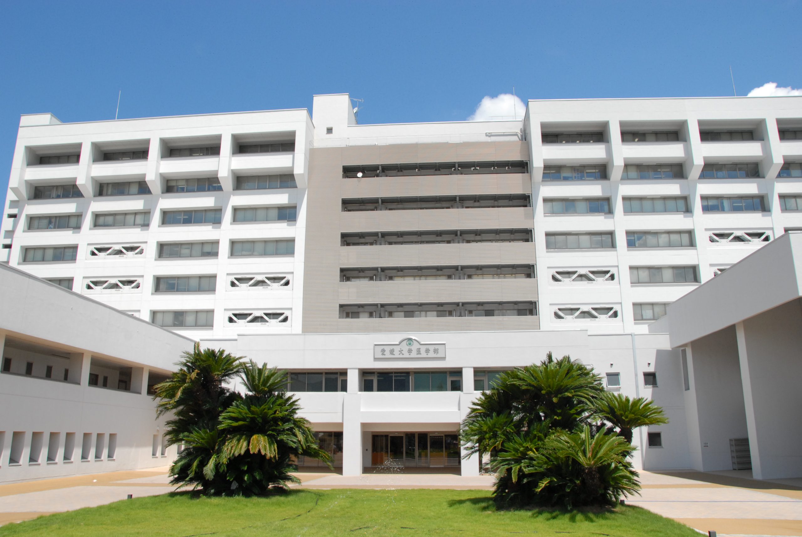
Course introduction

主な実績

  • Kouchi T, Tanabe Y, Smit EJ, et al. Clinical application of four-dimensional noise reduction filtering with a similarity algorithm in dynamic myocardial computed tomography perfusion imaging. Int J Cardiovasc Imaging. 2020 May 12. doi: 10.1007/s10554-020-01878-6.
  • Tanabe Y, Kido T, Kimura F, et al. Japanese Survey of Radiation Dose Associated With Coronary Computed Tomography Angiography - 2013 Data From a Multicenter Registry in Daily Practice. Circ J. 2020 Mar 25;84(4):601-608. doi: 10.1253/circj.CJ-19-0843.
  • Hirai K, Kido T, Kido T, et al. Feasibility of contrast-enhanced coronary artery magnetic resonance angiography using compressed sensing. J Cardiovasc Magn Reson. 2020 Feb 13;22(1):15. doi: 10.1186/s12968-020-0601-0.
  • Kawaguchi N, Okayama H, Kido T, et al. Clinical significance of corrected relative flow reserve derived from (13)N-ammonia positron emission tomography combined with coronary computed tomography angiography. J Nucl Cardiol. 2019 Nov 11. doi: 10.1007/s12350-019-01931-5.
  • Okada K, Matsuda M, Tsuda T, et al. Dual-energy computed tomography for evaluation of breast cancer: value of virtual monoenergetic images reconstructed with a noise-reduced monoenergetic reconstruction algorithm. Jpn J Radiol. 2020 Feb;38(2):154-164. doi: 10.1007/s11604-019-00897-1.
  • Tsuruoka S, Kataoka M, Hamamoto Y, et al. Tumor growth patterns on magnetic resonance imaging and treatment outcomes in patients with locally advanced cervical cancer treated with definitive radiotherapy. Int J Clin Oncol. 2019 Sep;24(9):1119-1128. doi: 10.1007/s10147-019-01457-3.
  • Kurata A, Fukuyama N, Hirai K, et al. On-Site Computed Tomography-Derived Fractional Flow Reserve Using a Machine-Learning Algorithm - Clinical Effectiveness in a Retrospective Multicenter Cohort. Circ J. 2019 Jun 25;83(7):1563-1571. doi: 10.1253/circj.CJ-19-0163.
  • Watanabe E, Miyagawa M, Uetani T, et al. Positron emission tomography/computed tomography detection of increased (18)F-fluorodeoxyglucose uptake in the cardiac atria of patients with atrial fibrillation. Int J Cardiol. 2019 May 15;283:171-177. doi: 10.1016/j.ijcard.2018.10.106.
  • Ogawa R, Kido T, Nakamura M, et al. Magnetic resonance assessment of fetal lung maturity: comparison between signal intensity and volume measurement. Jpn J Radiol. 2018 Jul;36(7):444-449. doi: 10.1007/s11604-018-0745-0.
  • Matsuda M, Tsuda T, Kido T, et al. Dual-Energy Computed Tomography in Patients With Small Hepatocellular Carcinoma: Utility of Noise-Reduced Monoenergetic Images for the Evaluation of Washout and Image Quality in the Equilibrium Phase. J Comput Assist Tomogr. 2018 Nov/Dec;42(6):937-943. doi: 10.1097/RCT.0000000000000752.

Staff introduction

スタッフ紹介

スタッフ紹介